
Fig. 2.
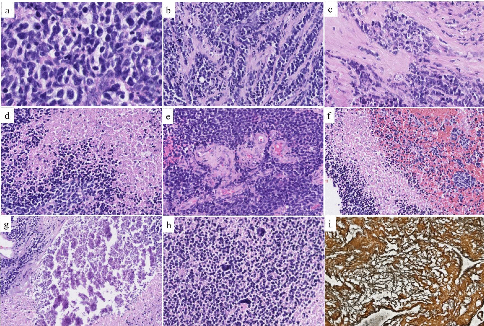
Histopathological features. a The tumor was highly cellular, composed of small cells with scant cytoplasm and hyperchromatic nuclei (HE, magnification 400×). b, c Intratumoral desmoplasia with focally tumoral cells disposed in a streaming pattern (HE, magnification 200×). d Tumoral geographical necrosis (HE, magnification 100×). e: Microvascular endothelial proliferation (HE, magnification 100×). f Intratumoral hemorrhage (HE, magnification 100×). g Calcifications (HE, magnification 200×). h Presence of numerous “bizarre cells” with high nuclear pleomorphism (HE, magnification 200×). i Reticulin stain highlighted intense desmoplasia (HE, magnification 200×). HE, Hematoxylin–eosin