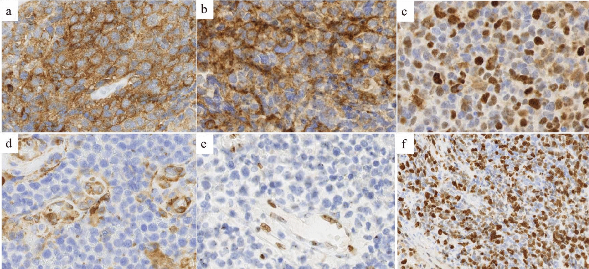
Fig. 3.

Immunohistochemical features. a Diffuse Synaptophysin immunopositivity (magnification 400×). b Diffuse CD56 immunopositivity (magnification 400×). c Intense nuclear immunopositivity of p53 protein in more than 10% of the tumoral cells (magnification 400×). d Loss of Filamine-A expression in tumoral cells with immunopositivity in endothelial cells (magnification 400×). e Loss of H3K27me3 expression in tumoral cells with immunopositivity in endothelial cells (magnification 400×). f High KI-67 proliferation index with some hotspot of more than 50% of immunopositivity in tumoral cells (magnification 200×)
