
Figure 1:
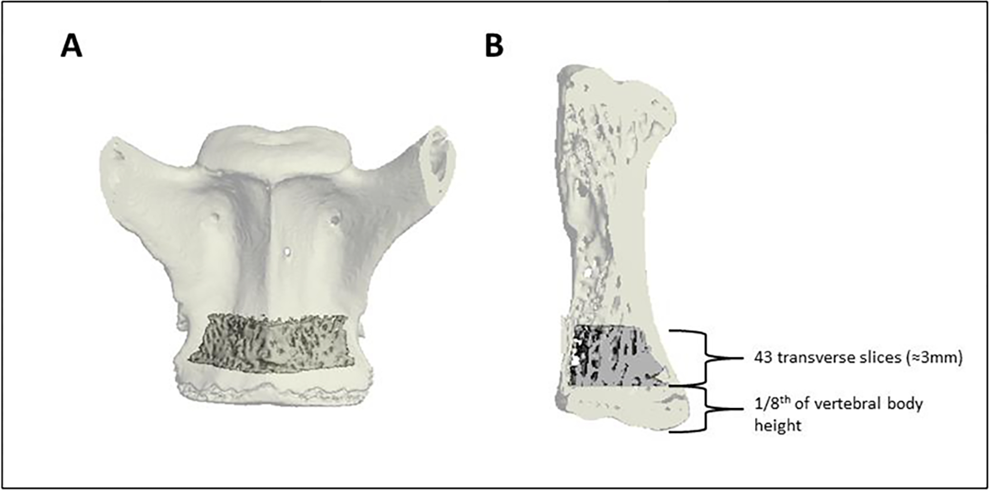
Coronal (A) and sagittal (B) views of the trabecular region of interest (ROI) in the caudal aspect of the more cranial of the two fused vertebrae. The trabecular region of interest began an eighth of the vertebral body height superior to the distal end of the vertebrae and extended cranially 43 transverse slices (≈3mm). The trabecular ROI is shown as solid within the transparent shell of the vertebrae.