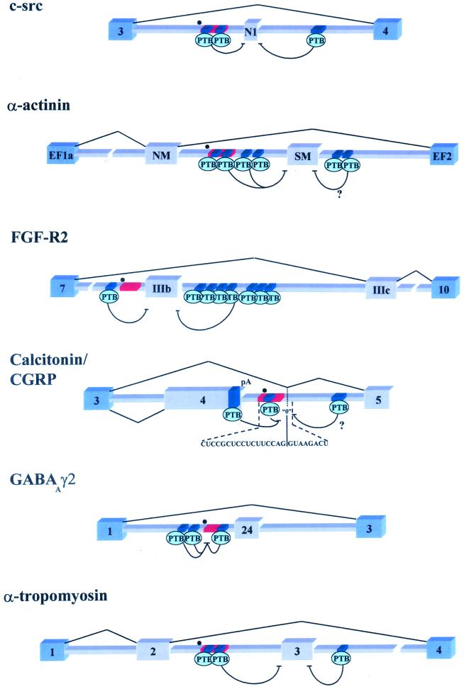
FIG. 2.
Schematic representation of six pre-mRNAs that are regulated by PTB through exon definition antagonism. Blue exons represent constitutively spliced exons, while the gray exons are regulated. Dark-blue segments represent PTB binding sites; red rectangles are the branch point-associated polypyrimidine tract, while the black dots represent identified or putative branch points. (c-src) The N1 exon is repressed in nonneural cell types by intronic PTB binding sites flanking the exon (9, 10, 12). (α-actinin) The SM exon may be repressed in tissues other than smooth muscle through a network of PTB binding sites located on both sides of the SM exon (65). It should be noted that in vitro the intron downstream of SM is not required for PTB repression. (FGF-R2) Exon IIIb is also repressed in mesenchymal tissues through multiple PTB binding sites found on either side of IIIb (8; E. J. Wagner and M. A. Garcia-Blanco, unpublished results). (Calcitonin/CGRP) The calcitonin-CGRP fourth exon is included and subsequently polyadenylated in the majority of tissues, possibly due to PTB-mediated repression of a zero-length exon located in the downstream intron. Repression of this exon blocks a potential recursive-splicing pathway to exon 5 (14, 39–42). Splicing to exon 5 occurs in neural cell types where PTB levels may be reduced. (GABAAγ2) The small 24-nucleotide exon is repressed in nonneural cell types through PTB binding near the branch point sequence (1, 72, 73). (α-tropomyosin). Exon 3 is specifically repressed in smooth muscle tissue by PTB or a novel PTB variant (25, 26, 55) (Smith, unpublished).

