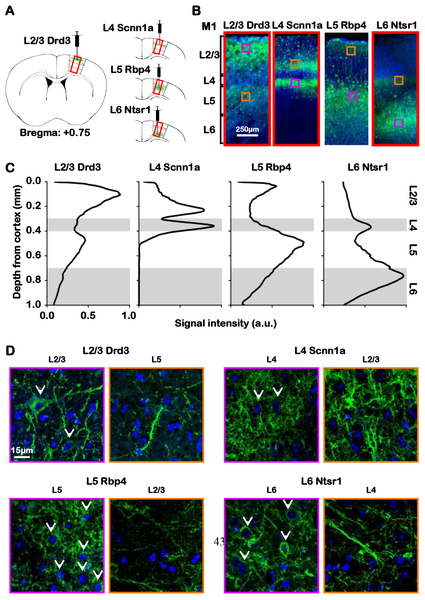
Figure 1: Histology reveals ChR2 expression of Drd3 L2/3, Scnn1a L4, Rbp4 L5 and Ntsr1 L6 Cre-line mice in their respective M1 layers and their projections.

(A) Illustration presents the viral injection sites in respective layers of M1. (B) Representative fluorescence images from the four layer-specific Cre-lines show robust ChR2-EYFP expression throughout their respective layers and intra-cortical projections in the M1. (C) Normalized signal intensity plots along the M1 cortical depth show a local peak throughout their respective layers and intra-cortical projections. (D) Merged representative confocal images of ChR2-EYFP and stained nuclear marker DAPI confirmed colocalization of ChR2-EYFP and M1 neurons throughout their respective layers (white arrows), but not their intra-cortical projections. Together, ChR2-EYFP expression is observed in L2/3 M1 neurons and L5 projections for Drd3 L2/3 Cre-line, in L4 M1 neurons and L2/3 projections for Scnn1a L4 Cre-line, in L5 M1 neurons and L2/3 projections for Rbp4 L5 Cre-line, and in L6 M1 neurons and L4 projections for Ntsr1 L6 Cre-line.
