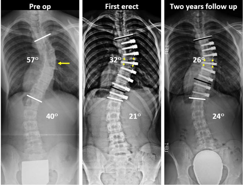
Figure 8.
Preoperative posteroanterior scoliosis radiograph of a mature group patient (Risser 4, proximal humerus ossification system 4 [PHOS 4]) with a 57° thoracic curve and 40° lumbar curve. First erect radiograph shows correction of major and compensatory curves. At 24 mo, the patient was Risser 5 (PHOS 5), and posteroanterior radiograph shows maintenance of clinically successful correction. Apical wedge angle was 12° at first erect and 8° at 2-y follow-up, calculating a VBSQ of 4°. Follow-up Cobb angles were measured between vertebra contained by the black bars, where white bars indicate preoperative angles. The yellow arrow indicates the apical vertebra, and yellow dots mark the vertebral body margins.

