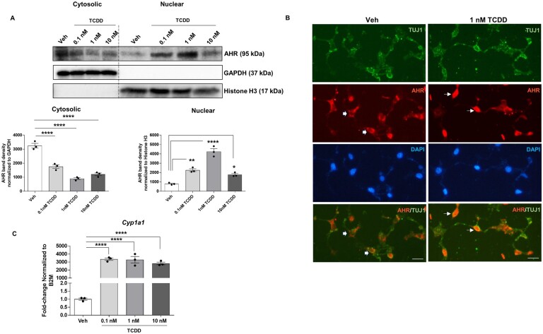
Figure 4.
TCDD induces translocation of AHR to the nucleus and target gene Cyp1a1 expression in IM-FEN cells. A, Western blot for AHR protein expression in the cytoplasmic and nuclear fractions of cells treated with TCDD (0.1, 1, and 10 nM) for 30 min, showing its nuclear translocation upon activation by TCDD. B, Representative images of AHR (red), TUJ1 (green), and DAPI (blue) immunostaining of IM-FEN cells treated with 1 nM TCDD for 24 h. Thick arrows point to cytoplasmic and line arrow point to nuclear (arrow) signals. C, Cyp1a1 gene expression in cells treated with various doses of TCDD (0.1, 1, and 10 nM) for 24 h by qPCR. Scale bar, 50 µm. Similar results were obtained in 3 independent experiments. Results are mean ± SEM, *p < .05; **p < .01; ****p < .0001.

