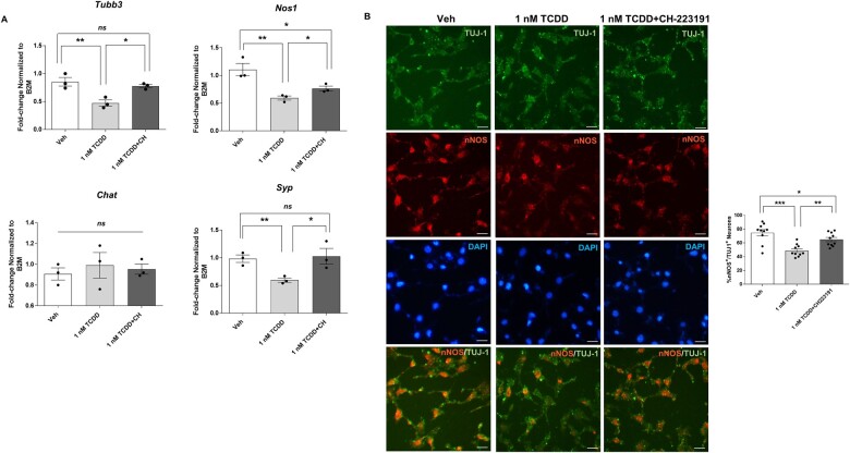
Figure 6.
Reduced expression of neuronal markers in IM-FEN cells exposed to TCDD is AHR dependent. Cells were pretreated with an AHR antagonist, CH-223191 (10 µM) for an hour and then treated with 1 nM TCDD for 24 h. A, Real-time PCR was done to assess the changes in Tubb3, Nos1, Chat, and Syp (synaptic marker). B, Representative images of neuronal marker TUJ-1 (green), nNOS (red), and DAPI (blue) immunostaining in IM-FEN cells treated with 1 nM TCDD in the presence or absence of 10 µM CH-223191. Data represent three independent experiments. Scale bar, 50 µm. Results are mean ± SEM, *p < .05; **p < .01; ***p < .001.

