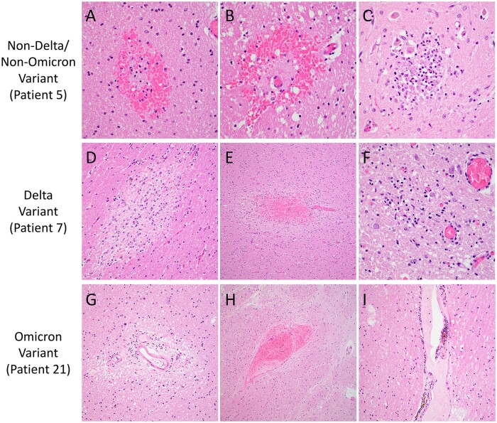
Figure 2.
Neuropathologic features of SARS-CoV-2 Delta, Omicron, and non-Delta/non-Omicron variant patients. Representative images show microhemorrhages in the occipital lobe (A) and basal ganglia (B) and a microinfarct in the medulla (C) of a non-Delta/Non-Omicron variant patient (Patient 5). A microinfarct in the midbrain (D), a microhemorrhage in the temporal lobe (E), and a microglial nodule in the medulla (F) are shown for a Delta variant patient (Patient 7). Perivascular rarefaction in the corpus callosum (G), perivascular microhemorrhage/red blood cell extravasation in the basal ganglia (H), and mild perivascular inflammation in the thalamus (I) are illustrated for an Omicron variant patient (Patient 21). Images taken with 40× objective (A–C, F), 20× (D, G, I), and 10× (E, H).

