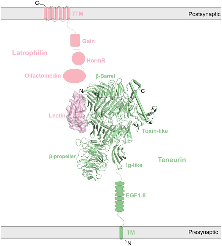
Figure 12.
Cryo-EM structure of 1:1 Teneurin (TEN2)/Latrophilin (LPHN3) complex (PDB: 6VHH). Four domains of TEN2 (Toxin-like, β-propeller, β-Barrel, and Ig-like) and one domain of LPHN3 (Lectin) are shown in the structure. The domains that are missing in the structure are shown as schematic representations. TEN2 (green, shown in cartoon representation) is localized to the presynaptic membrane, whereas LPHN3 (pink, shown in surface representation with semi-transparency) is localized to the postsynaptic membrane.

