
Figure 9: Acute HFD-induced activation of NMDA receptors is dependent upon activation of metabotropic glutamate and purinergic receptors.
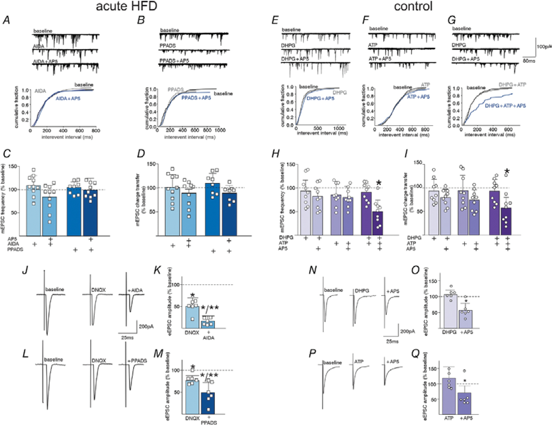
A. In an acute HFD DMV neuron, perfusion with the group I mGluR receptor antagonist, AIDA, prevented AP5 from decreasing mEPSC frequency (summarized by the cumulative fraction graph below). B. Similarly, perfusion with the P2X receptor antagonist, PPADS prevented the ability of AP5 to decease mEPSC frequency (summarized in the cumulative fraction graph below). C, D. Graphical summary of the effects of antagonists to block AP5 mediated effects on mEPSC frequency (C.) and charge transfer (D.) (N=10 neurons from 6 rats and N=8 neurons from 3 rats for AIDA and PPADS, respectively; two-way ANOVA with targeted contrasts: *p<0.05, one-way ANOVA with post-hoc Bonferroni multiple comparison test or two-tailed paired Student’s T-Test. Data are represented as mean±SD). E. In a control DMV neuron, perfusion with DHPG did not uncover any AP5-mediated decrease in mEPSC frequency (cumulative fraction graph below). F. Similarly, perfusion with the ATP did not uncover any AP5-mediated decrease in mEPSC frequency (cumulative fraction graph below). G. In contrast, perfusion both DHPG and ATP uncovered inhibitory effects of AP5 (cumulative fraction graph below). H. I. Graphical summary of the effects of agonists perfusion to uncover AP5 mediated inhibition of mEPSC frequency (H.) and charge transfer (I.) (DHPG, 3/9 neurons from 3 rats; ATP, 5/9 neurons from 3 rats; DHPG+ATP 8/9 neurons from 3 rats. Two-way ANOVA with targeted contrasts: *p<0.05, one-way ANOVA with post-hoc Bonferroni multiple comparison test or two-tailed paired Student’s T-Test. Data are represented as mean±SD). J. In an acute HFD DMV neuron, perfusion with DNQX was used to isolate an NMDA receptor mediated glutamate current, which was inhibited by subsequent perfusion with AIDA. K. Graphical summary of the effects of a AiDA to block NMDA receptor mediated eEPSCs. (N=6 neurons from 4 rats) L. Perfusion with DNQX decreased eEPSC amplitude and subsequent perfusion with PPADS inhibited the remaining evoked NMDA current. M. Graphical summary of the effects of PPADS to block NMDA receptor mediated eEPSCs (N=6 neurons from 3 rats). N. In a control DMV neuron, perfusion with DHPG uncovered the ability of AP5 to inhibit NMDA receptor mediated eEPSCs. O. Graphical summary of the effects of the DHPG, to uncover NMDA receptor mediated transmission in control DMV neurons (N=6 neurons from 3 rats; *p<0.05, two-tailed paired Student’s T-Test. Data are represented as mean±SD). P. In a control DMV neuron, perfusion with ATP uncovered the ability of AP5 to inhibit NMDA receptor mediated eEPSCs. Q. Graphical summary of the effects of ATP, to uncover NMDA receptor mediated transmission in control DMV neurons. (N=6 neurons from 3 rats) *p<0.05, two-tailed paired Student’s T-Test. Data are represented as mean±SD