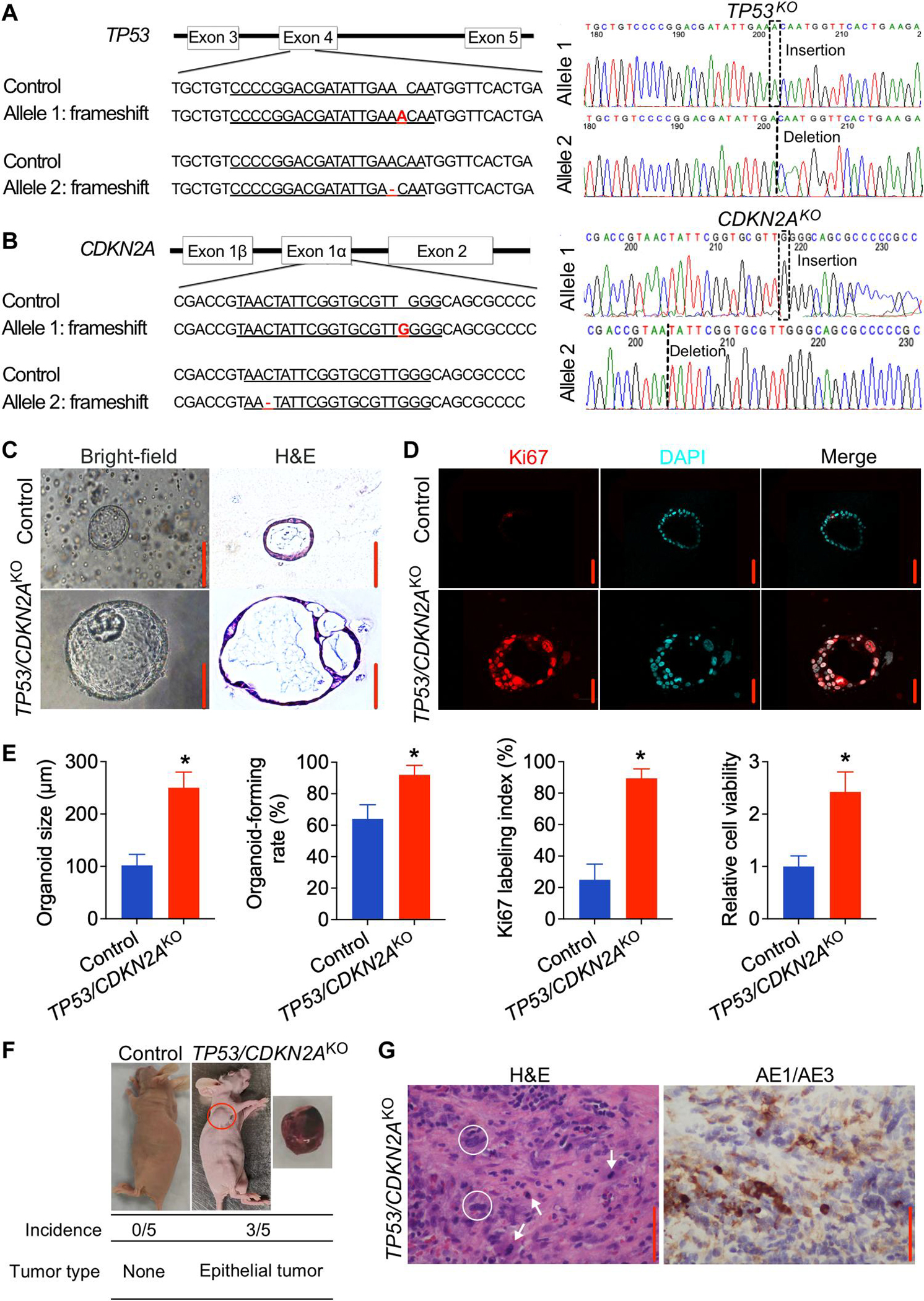
Fig. 2. Knockout of TP53/CDKN2A promotes neoplastic transformation in human normal GEJ organoids.

(A and B) Sanger sequencing of TP53/CDKN2AKO GEJ organoids showing 1-bp insertion or deletion in TP53 (A) or CDKN2A (B). Red font indicates corresponding frameshift indels in the genomic DNA. (C and D) On the 10th day after seeding 1 × 10⁵ dissociated organoid cells, organoid cultures were photomicrographed using phase-contrast microscopy and collected for (C) bright-field, H&E, and (D) IF staining for Ki67 (red color). DAPI, 4′,6-diamidino-2-phenylindole. (E) Average organoid size, organoid-forming efficiency, and Ki67 index were determined by measuring >50 organoids. Data are represented as means ± SD; n = 4 biologic replicates. *P < 0.05 by Student’s t test. (F) Representative images of xenografts from mice injected with control or TP53/CDKN2AKO GEJ organoids; the underlying table shows incidence and tumor characteristics. This experiment was repeated once with similar results. (G) Representative H&E and AE1/AE3 pan-keratin IHC staining (brown) in xenografts arising from TP53/CDKN2AKO organoids. White arrows, mitoses; white circles, abnormally large, pleomorphic cells with irregular nuclear envelopes; scale bars, 100 μm.
