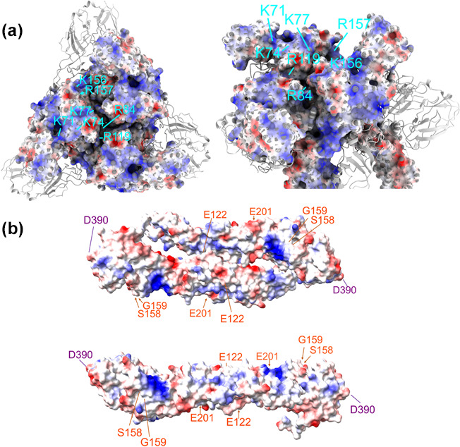
Fig. 3.

(a) Top and rotated and cropped three-dimensional model of the mature trimeric spike complex of EEEV E1 and E2 heterodimers (PDB: 6M×4). E1 is shown as a ribbon diagram and E2 as a residue electrostatic map (red=negative charge and blue=positive charge). E2 residues K71/K74/K77, R84/R119, K156/R157 (identified with lines in cyan) have been shown to be three separate interaction sites involved in HS binding by naturally circulating strains. (b) Top and side view of an electrostatic three-dimensional model of the JEV E dimer ectodomain (PDB: 5MV1, dimer was reconstructed using Swiss model programming). Corresponding residues on both E proteins where mutations increase HS interactions for TBEV (E122, S158, G159 and E201) are highlighted in orange, and for MVEV (D390) is highlighted in purple. The figures were made using UCSF ChimeraX software.
