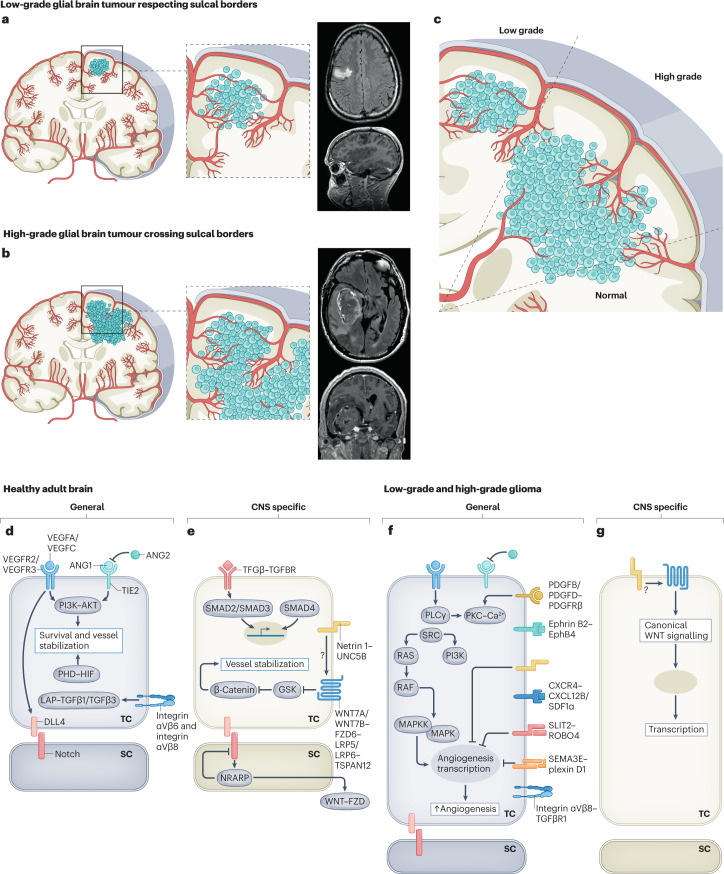
Fig. 5. Molecular mechanisms regulating the vasculature during initiation and progression of glial brain tumours.
Figure illustrating the hypothetical concept postulating the gyral confinement and respect of sulcal borders during progression from low-grade glial brain tumours to high grade glial brain tumours based on radiological observations and the concept of sprouting angiogenesis and recruitment of blood vessels from the adjacent brain parenchyma. a–c, Cross sections of the adult human brain in the coronal plane showing pathological angiogenesis in glial brain tumours. Illustrations and T1-weighted coronal and sagittal MRI scans with gadolinium show that low-grade gliomas are often confined to one gyrus, thereby respecting sulcal borders (parts a,c). Illustrations and T1-weighted coronal and sagittal MRI scans with gadolinium show that invasive high-grade gliomas do often not respect gyral confinement and cross sulcal borders (parts b,c). d,e, Molecularly, numerous signalling pathways have been implicated in the adult healthy brain, regulating endothelial cell quiescence, survival and maintained inhibition of paracellular permeability. Molecular cues can be either non-CNS specific (part d) or CNS specific (part e). These signalling pathways are thought to be of importance during both embryonic and postnatal vascular brain development, as well as to contribute to the maintenance of the quiescent healthy adult brain vasculature. f,g, Molecularly, different non-CNS-specific and CNS-specific angiogenic molecular mechanisms have been implicated in glioma initiation and progression, and they include the reactivation of developmentally active ligand–receptor pairs. ANG1, angiopoietin 1; ANG2, angiopoietin 2; SC, stalk cell; TC, tip cell. Images in parts a,b courtesy of P. Nicholson.

