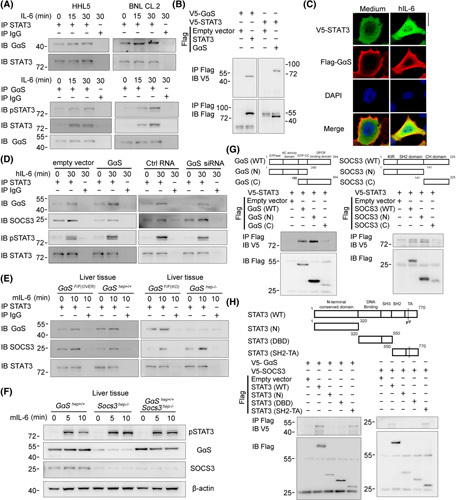
FIGURE 4.

Cytoplasm‐translocated GαS associates with STAT3 upon IL‐6 stimulation to impede SOCS3–STAT3 interaction. (A) The association between GαS and STAT3 was examined by co‐IP in HHL5 and BNL CL.2 cells treated with IL‐6 for the indicated time periods. (B) Tagged STAT3 and GαS constructs were cotransfected into HEK293T cells as indicated, and the association between GαS and STAT3 was examined by co‐IP. (C) V5‐tagged STAT3 and Flag‐tagged GαS constructs were cotransfected into HHL5 cells. Confocal microscopic images of these cells upon IL‐6 stimulation are shown as indicated. Scale bar, 10 μm. (D) HHL5 cells were transfected with control, GαS overexpression plasmids, or GαS small interfering RNA as indicated; the associations between STAT3 and GαS or SOCS3, respectively, were analyzed by co‐IP. (E) Male GαSF/F(OVER) and GαShep+/+ , GαSF/F(KO) and GαShep−/− mice were injected with recombinant IL‐6 through the hepatic portal vein; the associations between STAT3 and GαS or SOCS3, respectively, were analyzed by co‐IP in liver tissue lysates. (F) Male GαShep+/+ , Socs3hep−/− , and GαShep+/+Socs3hep−/− mice were injected with recombinant IL‐6 through the hepatic portal vein for the indicated time periods, and STAT3 phosphorylation was examined by western blot in the liver tissues. (G,H) Tagged STAT3, GαS, SOCS3 and their truncates were constructed and cotransfected into HEK293T cells, and cell lysates were precipitated with Flag antibody and immunoblotted with V5 antibody as indicated. Data are shown as photographs from one representative of three independent experiments. Abbreviations: Ctrl, control; DBD, DNA binding domain; h‐, human; IB, immunoblotting; IP, immunoprecipitation; m‐, mouse; p‐, phosphorylated; SH2‐TA, Src homology 2‐transactivation; WT, wild type
