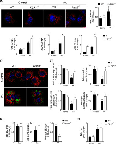
FIGURE 4.

Ripk3 deficiency dampens hepatocyte mitochondrial reactive oxygen species (ROS) following palmitate treatment in AML‐12 cells. Wild‐type (WT) and Ripk3 −/− AML‐12 cells were treated with 125 μM palmitate (PA) for 24 h. (A) Representative staining for MitoSOX Red (red). Nuclei were counterstained with Hoechst 33258 (blue). Scale bar = 85 μM. Histogram shows the quantification of mitochondrial ROS. (B) qRT‐PCR analysis of Nfr1, Sod2, and Srit3 in AML‐12 cells. (C) Representative staining for tetramethylrhodamine methyl ester perchlorate (TMRM) (red), LipidTOX Green (green), and Hoechst 33342 (blue) for mitochondrial network, neutral LD, mitochondrial superoxide, and nuclei, respectively. Scale bar = 85 μM. (D) Histograms show the quantification of TMRM intensity and number, area, and average size of mitochondria. (E) Histograms show the quantification of total area, average size, and number of lipid droplets. (F) Fluorometric measurement of Nile red staining normalized by SRB method. Results are expressed as mean ± SEM fold change or percentage of four independent cultures from each genotype § p < 0.05 and *p < 0.01 from control; † p < 0.05 from and ‡ p < 0.01 from respective control.
