Figure 3.
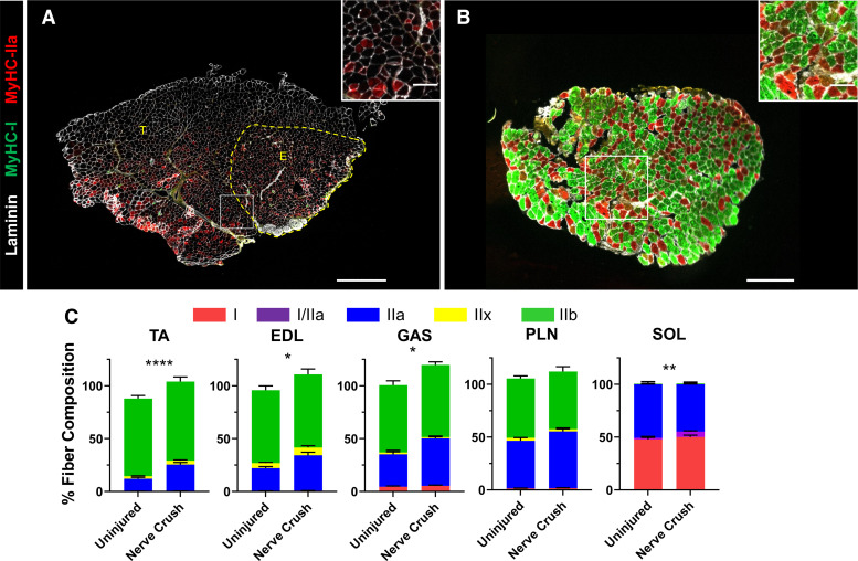
I/IIa hybrid and IIa myofibers increase in aged ephrin-A3−/− mice after nerve crush. Immunohistochemistry showing laminin (white), myosin heavy chain (MyHC)-I (green), and MyHC-IIa (red) expression in transverse cryosections from tibialis anterior (TA)-extensor digitorum longus (EDL) (A) and soleus (SOL) muscles from aged ephin-A3−/− mice 4 wk post-nerve crush (B). C: fiber-type distribution of the TA, EDL, gastrocnemius (GAS), plantaris (PLN), and SOL muscles of aged ephrin-A3−/− mice. Data are presented as means ± SE. *P < 0.05; **P < 0.01; ****P < 0.0001. A: scale bar, 600 µm. B: scale bar, 300 µm. Inset scale bar, 100 µm.

