
Fig. 7.
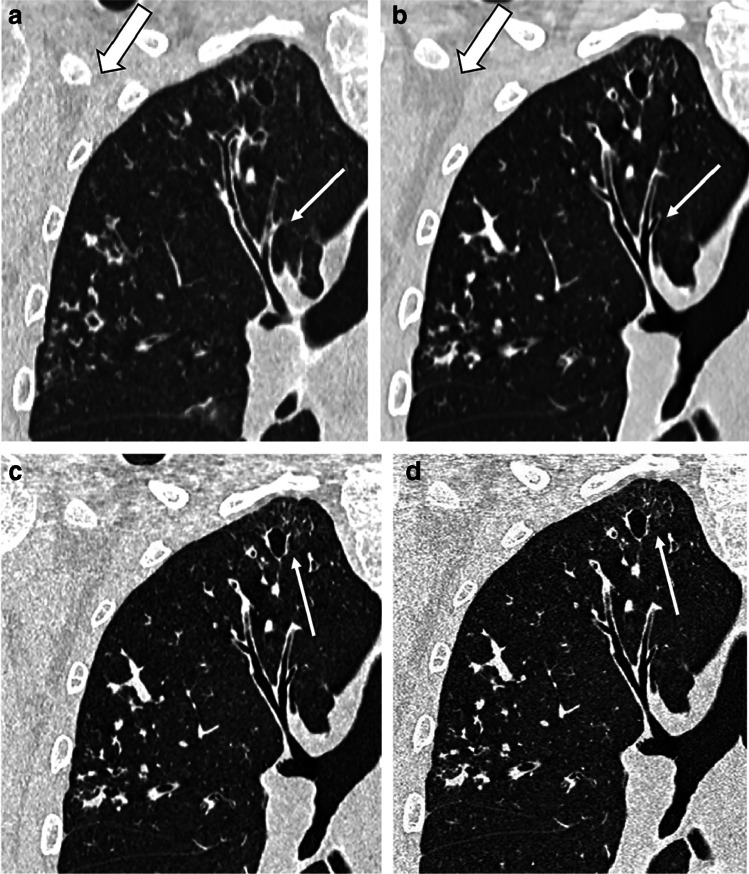
Photon-counting detector CT (PCD-CT) and conventional energy integrating detector CT (EID-CT) scanning in a 20-year-old man with cystic fibrosis. a–d Coronal detail of end-inspiratory CT using conventional EID-CT (a) and PCD-CT (b–d). Images (a) and (b) have slice thickness of 1 mm, and images (c) and (d), 0.6 mm and 0.2 mm, respectively. All four images have similar kernels including iterative reconstruction. Comparison of images (a) and (b) shows a clear reduction of image noise (reduced granularity in the axillary region, thick arrow) and sharper definition of bronchial wall and cystic bronchiectasis in the PCD-CT (thin arrow). In images (c) and (d), the increased resolution allows sharper details of the most peripheral structures (thin arrow)