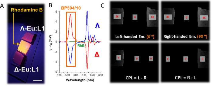
Fig. 5. Solid state time-resolved EDCC photography of an organic emitter and a CPL active Eu(III) complex.
A Conventional photo of rhodamine B (RhB) and Λ- and Δ -Eu:L1 in embedded into a PMMA matrix (C = 3 × 10⁻⁶ M) using 365 nm UV illumination. Scale bar = 1 cm. B CPL emission spectra of (green) rhodamine B, Δ- (red) and Λ- (blue) enantiomers of Eu:L1 in PMMA (λex = 365 nm) highlighting the spectral window selected for photography using an BP594/10 (OD4.0) filter. C Time-resolved (td = 20 µs) Images extracted from the quad polarisation view camera highlighting the recorded total emission, right- and left-handed emission with respect to the built-in polariser orientation to the fixed QWP fast axis. Numbers in red are avg. 8-bit pixel intensity values for each image region, tacq. = 400 ms, 10 avg. image, 100 total image accumulation.

