
Fig. 2.
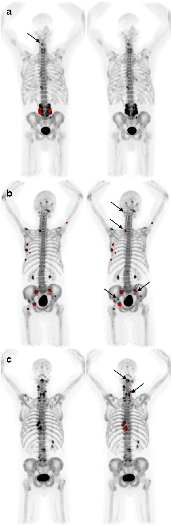
a Left: the AI model detects suspected a metastatic uptake in the lumbar spine and a small lesion in the cervical spine (arrow), marked in red. PET index 0.57%. Right: none of the physicians identify any lesions in the same patient. Of note, the patient had previously undergone lumbar spinal fusion. b Left: the AI model identifies several foci in the right ribcage, pelvis, and right hip. PET index 1.34%. Right: reader D identifies additional lesions in the skull, spine, and pelvis (arrows). PET index 1.42%. c Left: the AI model and the readers do not detect any lesions. Right: the threshold marks several lesions in the cervical (arrows) and thoracic spine. PET index 0.16%