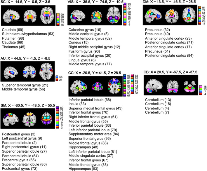
FIGURE 1.

Resting state networks (RSNs). Spatial maps of the RSNs (n = 53) derived from a spatially constrained group independent component analysis are plotted at the exhibited sagittal, coronal, and axial slices. The RSNs are partitioned into brain sub‐domains based on anatomical and functional properties of the (n = 53) brain components: AUD, Auditory; CC, Cognitive control; CEREB, Cerebellum; DMN, Default mode network; SC, Subcortical; SM, Somatomotor; VIS, Visual
