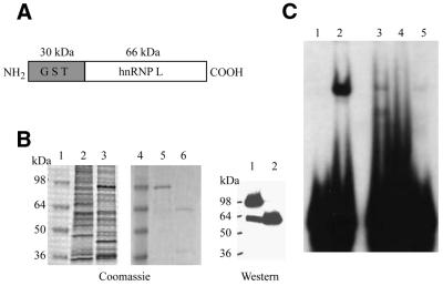
Figure 4.
Reconstitution of the nCaRE-B2-binding complex and peptide interference assay. (A) Domain organization of the GST–hnRNP-L fusion protein. (B) Purification of recombinant hnRNP-L protein from Sf9 cells. (Left) Coomassie brilliant blue staining: lanes 1 and 4, protein standards; lane 2, uninfected Sf9 extract; lane 3, Sf9 extract expressing GST–hnRNP-L; lanes 5 and 6, purified GST–hnRNP-L before (lane 5) and after (lane 6) cleavage with thrombin. (Right) Western analysis of the proteins in lanes 5 and 6 with anti-hnRNP-L antibody. (C) In vitro reconstitution of nCaRE-B2-binding activity by EMSA. Lane 1, no protein; lane 2, HeLa nuclear extract; lane 3, APE1 and hnRNP-L; lane 4, APE1 alone; lane 5, hnRNP-L alone.

