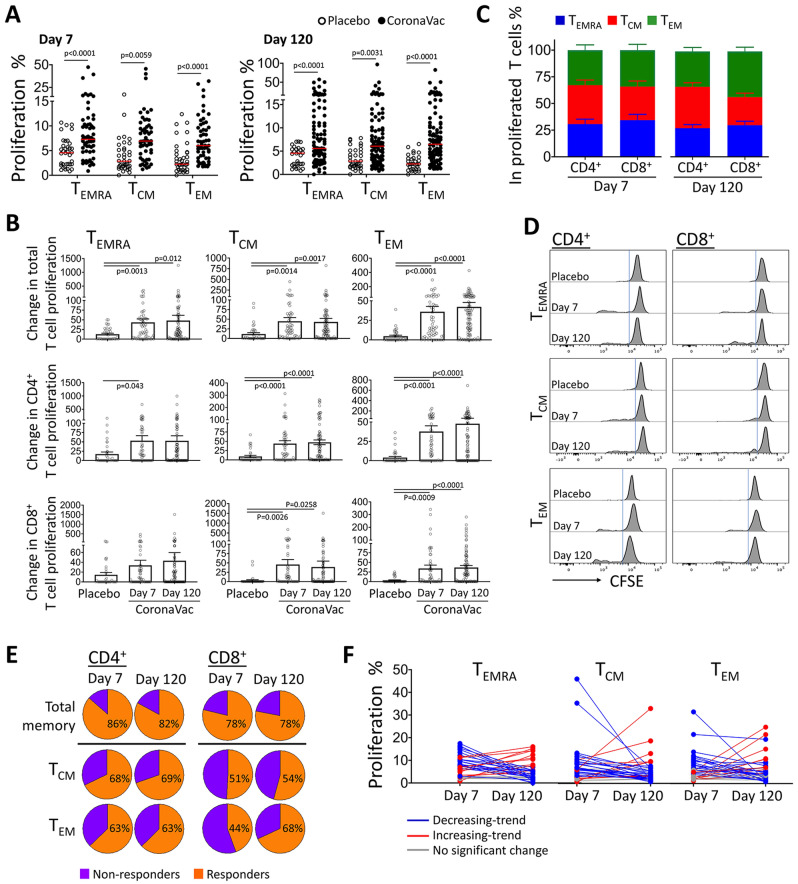
Figure 1.
Proliferation responses in memory T cells from the individuals after the aluminum-adjuvanted inactivated whole-virion SARS-CoV-2 vaccination demonstrated a considerable response against S1 antigen. Autologous monocyte-derived dendritic cells were loaded with SARS-CoV-2 Spike Glycoprotein-S1 and co-cultured with terminally-differentiated effector T (TEMRA), central memory T (TCM), and effector memory T (TEM) cells purified from the individuals after the second dose of CoronaVac (n = 48 on day 7 and n = 81 on day 120) or placebo administration (n = 34) on day 7 and day 120. Data obtained from the experiments without any technical complications are plotted; therefore, the number of volunteers for each group do not match to those of the total number of participants enrolled to the study. (A) Percentage of proliferation in memory T cell subsets is shown for each case. (B) Change in CD4+ and CD8+ memory T cell proliferation was calculated in comparison to the T cells co-cultured with non-specific antigen-loaded mDCs (without S1 antigen loading). (C) Contribution of each memory T cell subset to the overall proliferation response in CD4+ and CD8+ cells. (D) Flow cytometry histograms displaying representative proliferation responses from T cell subtypes are shown. (E) The percentage of participants whose T cells were responded to S1-loaded mDCs and proliferated more than the median value of the placebo group is demonstrated as “responders”. (F) The proliferation of TEMRA, TCM, and TEM obtained on day 7 and day 120 is given on individual basis (n = 32). (Significance was determined by Mann–Whitney U test for (A) and One-Way ANOVA for (B), *p < 0.05, **p < 0.01). Each dot represents a single participant.

