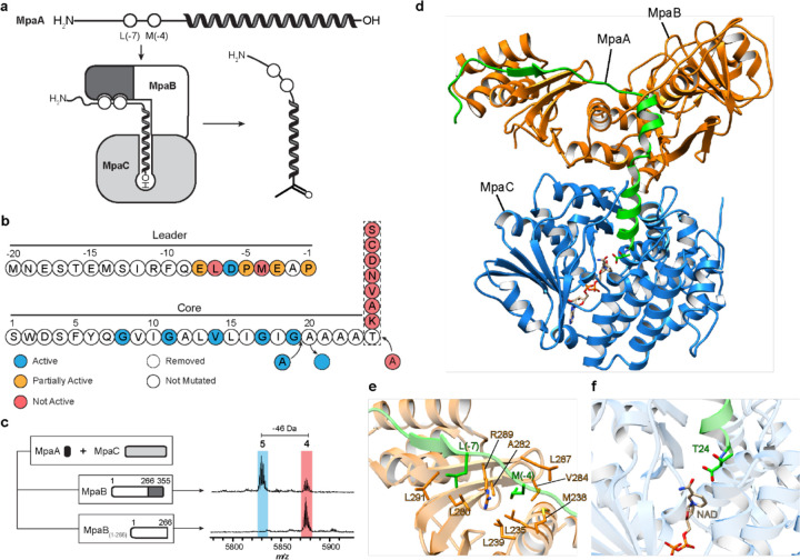
Fig. 4: Characterization of ketone intermediate formation by MpaB and MpaC.
a, Proposed interaction model for MpaA, MpaB, and MpaC. The RRE domain (dark grey) of MpaB interacts with the conserved leader region of MpaA. The C-terminus (depicted as “-OH”) of MpaA is directed into the active site of MpaC for modification. b, Summary of MpaA variant processing upon E. coli co-expression with MpaB/C. c, Mass spectra of MpaA after co-expression with MpaC, and MpaB or MpaB1–266. The RRE domain of MpaB is dark grey. d, The structure of MpaA1-MpaB-MpaC was predicted using AlphaFold-Multimer40. MpaA1, MpaB and MpaC are shown as cartoons in green, orange, and blue, respectively. The positioning of cofactor NADP was imported using a homologous alcohol dehydrogenase (PDB ID: 6C76) as a template. e, View of the leader peptide-RRE domain binding interface40. Select residues of MpaA and MpaB are in green and orange, respectively. f, View of the C-terminal Thr24 of MpaA in the substrate-binding pocket of MpaC.

