Figure 10.


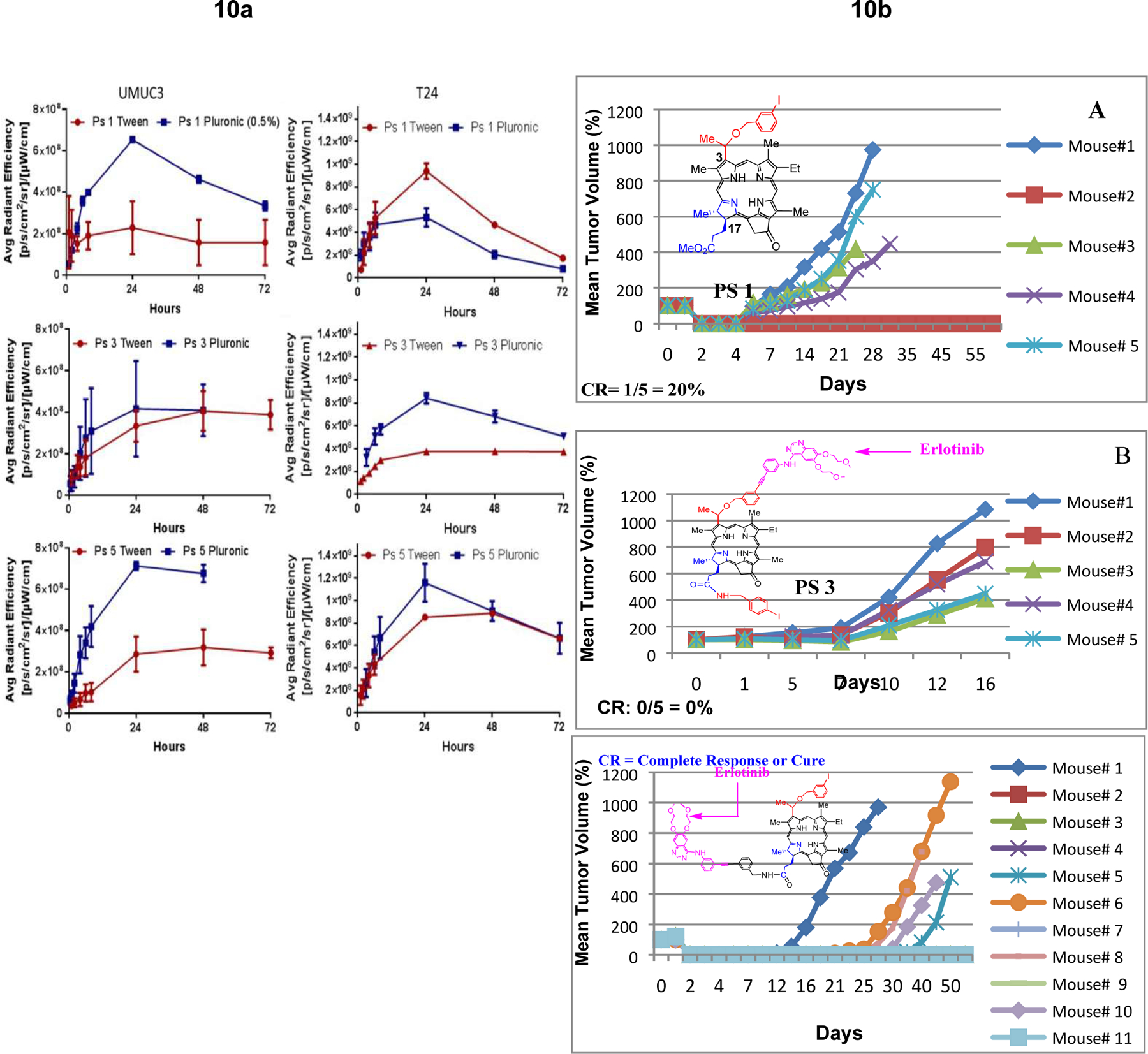
a: Comparative uptake of PS 1, 3, 5 (formulated either in Tween or Pluronic) in tumor, liver and skin (SCID mice bearing UMUC3 and T24 tumors) at a dose of 0.47 μmol/kg)) at variable time points (λex: 675 nm, λem: 720), using the IVIS optical imaging system.
b: Comparative in vivo PDT efficacy (long-term antitumor activity) of PS with and without erlotinib conjugates. (A): PS 1 (non-erlotinib PS), (B): PS 3 erlotinib moiety is attached at position-3 of the PS (C): PS 5, erlotinib moiety is attached at position-17 of the PS. PS 1, 3, and 5 were individually injected (dose: 0.47 micromole/Kg) to SCID mice bearing UMUC-3 tumors at the flank. Mice were exposed to light (665 nm, 135 J/cm2, 75 mW/cm2) at 24 h post-injection (optimal uptake time) and tumor re-growth was monitored daily. These results suggest that position of the erlotinib moiety in the PS makes a remarkable difference in long-term tumor cure.
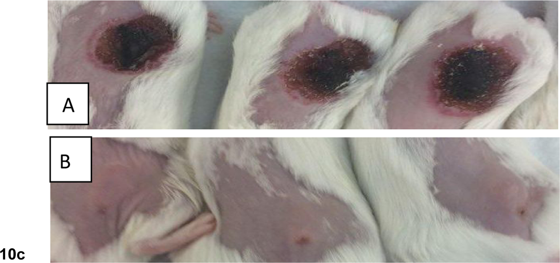
c: Comparative tumor necrosis of mice injected with (A) PS 3 (effective PDT agent), and (B) PS 5 that showed limited efficacy at 72h post-light exposure. Mice were treated under similar drug and light doses. Dug dose: 0.47 μmol/kg, light dose: 135 J/cm2, 75 mW/cm2 and the tumors were exposed to light (665 nm) at 24h post-injection of the PS.
