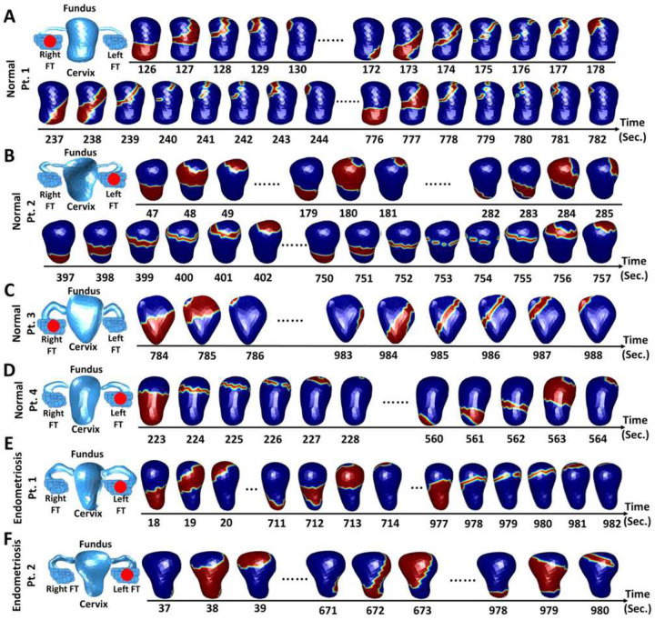
Figure 5.
Representative asymmetric uterine peristalsis patterns in healthy participants with the normal menstrual cycle (A-D) and endometriosis patients (E-F) during the ovulatory phase. In each panel, anatomical uterus geometry with fallopian tubes was segmented from the T1-weighted and T2-weighted MRI images. Red dots indicate the ovary with the dominant follicle. (A, C) Normal patients 1 and 3 have left-dominant follicles and left-sided asymmetric uterine peristalsis propagation. (B, D) Normal participants 2 and 4 have right-dominant follicles and right-sided asymmetric uterine peristalsis propagation. (E, F) Endometriosis patients with left dominant follicles and right-sided asymmetric uterine peristalsis propagation. Patient numbers correspond with data in Table 1

