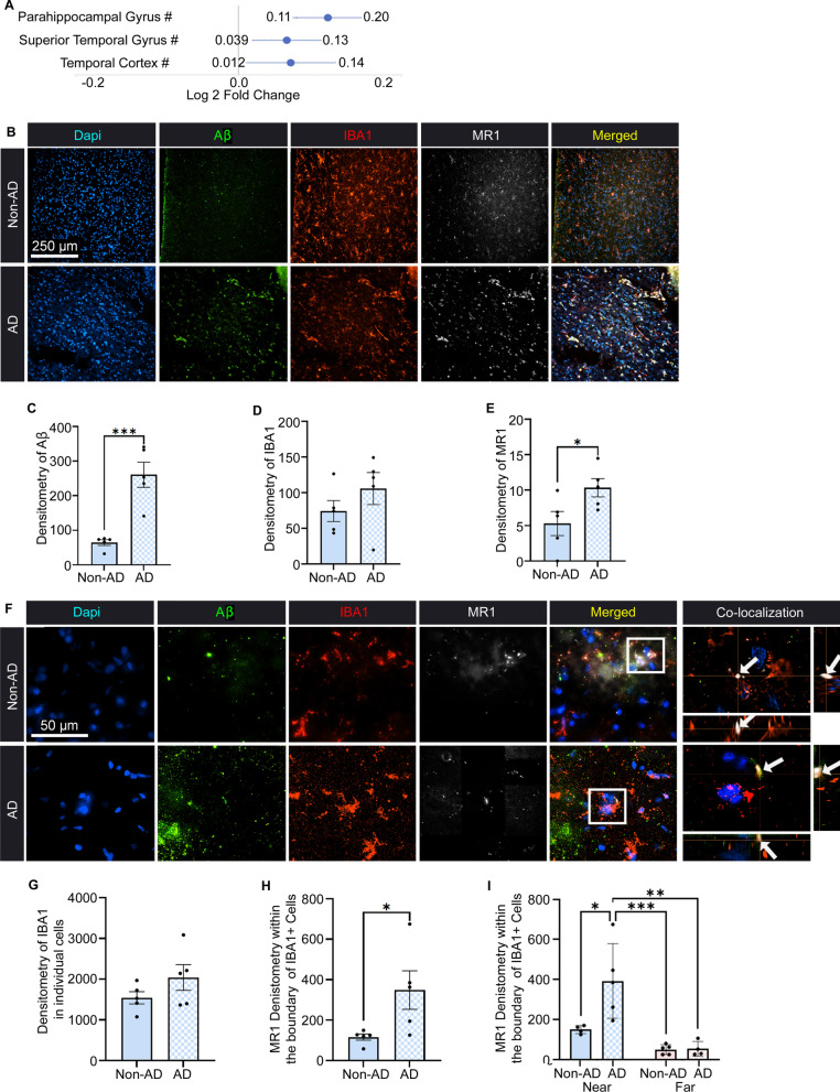
Fig. 1.
Immunostaining of microglia, amyloid-beta (Aβ), and MR1 in the temporal cortex of individuals with Alzheimer’s disease (AD). A Fold change in MR1 gene expression in human AD from the Agora Database. B Representative images of AD and non-AD control tissue showing Dapi (blue), Aβ (green), IBA1-labeled microglia/macrophages (red), MR1 (white), and these images merged. Scale bar = 250 µm. C–E Densitometry analysis presented as the relative mean pixel intensity for Aβ (C), IBA1 (D), and MR1 (E). F Representative images of non-AD and AD tissue at 100 × with co-localization of the boxed regions in the XY, ZX, and ZY planes (arrows). Scale bar = 50 µm. G, H Densitometry analysis presented as the relative mean pixel intensity for IBA1 (G) and MR1 expression within the boundaries of the IBA1 + cells (H). I Densitometry analysis of the MR1 signal within the boundaries of the IBA1 + cells that are determined to be near (visibly touching) or far from Aβ plaques (no visible touching). Statistical analysis was performed using a Student’s t test (C–E, G–I) and a two-way ANOVA with Tukey post hoc multiple comparison test (I). *P < 0.05, **P < 0.01, ***P < 0.001 (n = 5/group). Significance in both males and females was normalized to the age of death. Data are shown as the mean ± standard error of the mean

