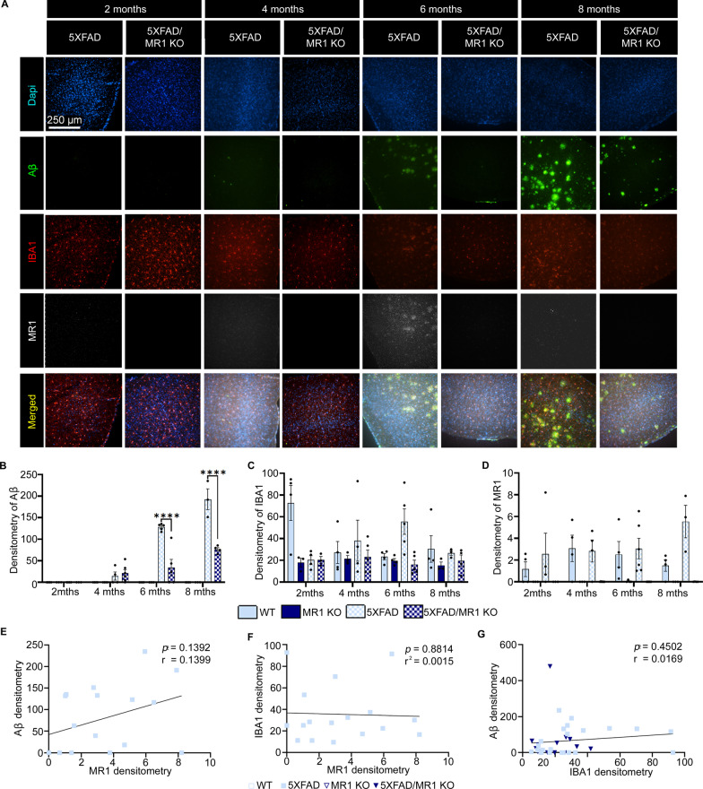
Fig. 2.
Densitometry analysis of microglia, amyloid-beta (Aβ) plaques, and MR1 in the temporal cortex of 5XFAD mice. A Representative images highlighting amyloid beta pathology showing Dapi (blue), IBA1-labeled microglia/macrophages (red), Aβ (green), MR1 (white), and the images merged. Scale bar = 250 µm. B–D Densitometry analysis as the relative mean pixel intensity for Aβ (B), IBA1 (C), and MR1 (D). E–G Graphs for the correlational analysis of the densitometry results between MR1 and Aβ compare 5XFAD (squares) and 5XFAD/MR1 KO mice (upside-down triangles) (E), MR1 and IBA1 (F), and IBA1 and Aβ (G). Statistical analysis was performed using a two-way ANOVA with Tukey post hoc multiple comparison test (B–D) or a simple linear regression analysis (E–G). ****P < 0.0001 (B–D: n = 3–7/group; E–G: n = 17–21/group). The data are shown as the mean ± standard error of the mean

