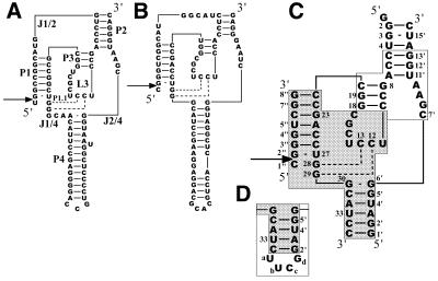
Figure 1.
Nucleotide sequences and possible secondary structures of HDV ribozymes. (A) Genomic HDV ribozyme. The stem, loop and junction regions are labeled according to the notations of Been (7). (B) Antigenomic HDV ribozyme. (C) Sequence and numbering for Rz-3, which consists of three RNA oligomer strands. The ribozyme contains a hybrid sequence of the genomic sequence (shown in a white box) and antigenomic sequence (shown in a shaded box). (D) Partial sequence (P4 region) of Rz-2, which consists of two RNA oligomer strands, and its numbering. Arrows indicate the cleavage site.

