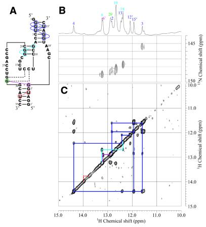
Figure 4.
Secondary structure (A), 15N-1H HSQC (B) and NOESY (C) spectra of the enzyme part (Ea35:Eb15) for Rz-3 measured in 10 mM sodium phosphate buffer, pH 6.5, 50 mM NaCl containing 5% D2O at 25°C. (B) The sample contained a mixture of 13C,15N-G-labeled Ea35 and non-labeled Eb16 (0.3 mM each). (C) The sample contained a mixture of non-labeled Ea35 and Eb16 (1 mM each). Blue, light blue and purple lines indicate NOE connectivities for P2 and P3 and the NOE between G29 and G6′, respectively. Dotted red lines indicate the putative NOE between U33 and U4′ in P4, which is expected from the assignment for Rz-2. The one-dimensional 1H spectrum and signal assignments are shown at the top.

