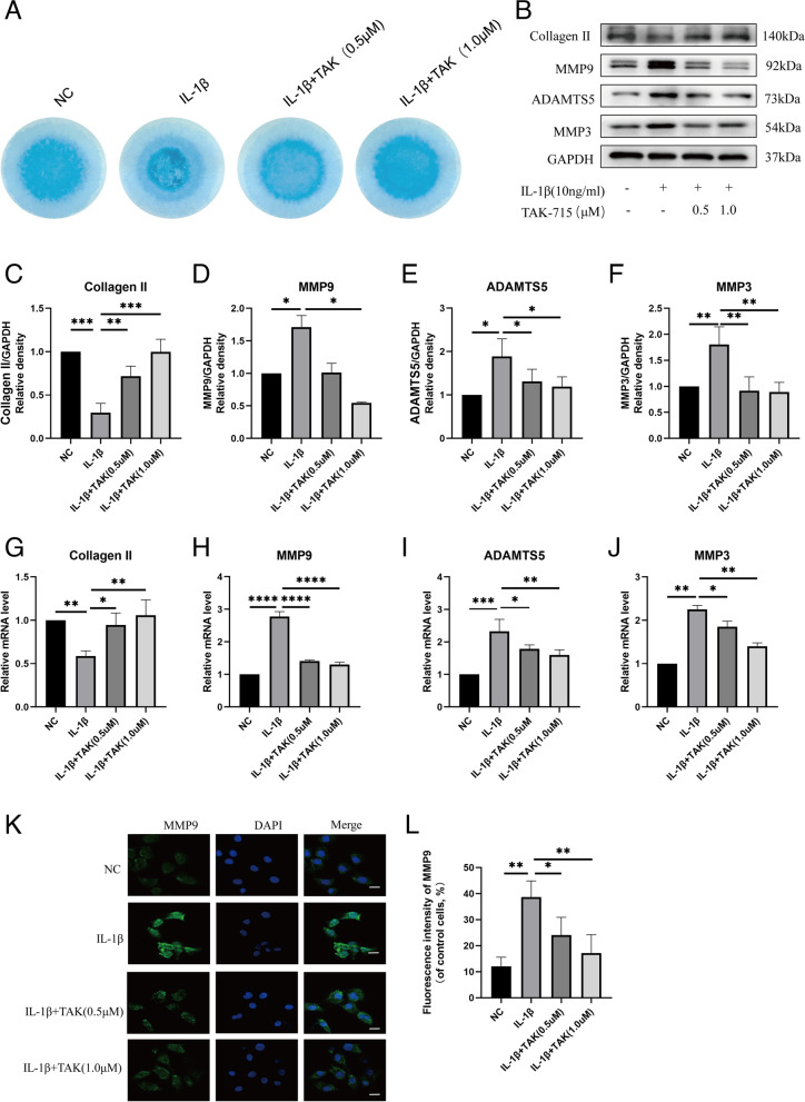
Fig. 2.
TAK-715 suppressed IL-1β-induced ECM degradation in NPCs. A NP cells were seeded in 24-well plates at 10.7/ml, which were in a 2D system. NP cells were co-treated with TAK-715 and IL-1β for 5 days, and then stained with alcian blue. B Western blot analysis was used to analyze Collagen II, MMP9, ADAMTS5 and MMP3 in NPCs; C-F The immunoblots of Collagen II, MMP9, ADAMTS5 and MMP3 were quantitatively analyzed. G-J The mRNA expression of Collagen II, MMP9, ADAMTS5 and MMP3 was measured by qRT‒PCR. K The fluorescence intensity of MMP9 was analyzed by immunofluorescence assays (scale bar:20 μm). J ImageJ was used to analyze the fluorescence intensity of MMP9. The data are presented as the mean ± SD (*P < 0.05; **P < 0.01; ***P < 0.001; ****P < 0.0001)

