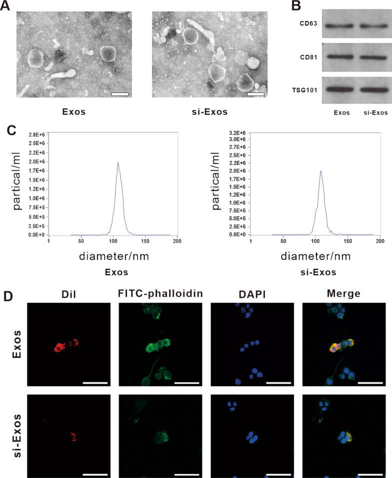
Fig. 1.
Characterization and internalization of HUVECs-derived Exos by RAW264.7. A Morphology of Exos and si-Exos identified by TEM. Scale bar = 200 nm. B Western blot analysis of the specific markers of exosomes, including CD63, CD81 and TSG101. C The particle size distribution and particle concentration of Exos and si-Exos detected by NTA. D The uptake of Exos and si-Exos by RAW264.7 cells. FITC-phalloidin (green) and DAPI (blue) were used to stain the cytoskeleton and nucleus of RAW264.7, respectively. Exosomes were labeled with Dil (red). Scale bar = 50 μm

