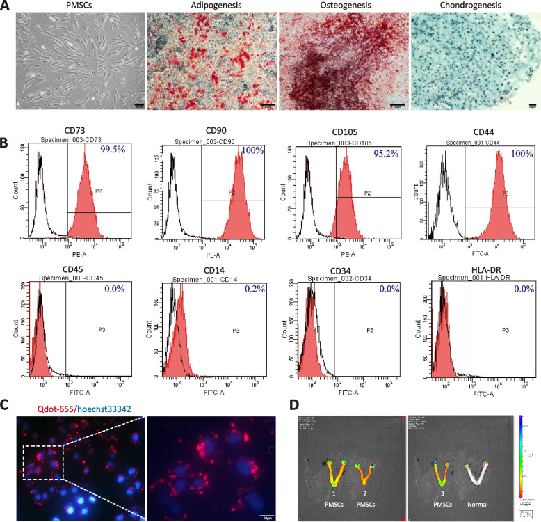
Fig. 1.
Characterization and identification of human PMSCs. A Morphology of PMSCs at the fourth passage. Oil Red O staining was conducted for adipogenic differentiation, alizarin red staining was used for osteogenic identification, and Alcian blue indicated chondrogenesis. Scale bar: 50 or 200 µm. B human PMSCs were positive for CD73, CD90, CD105, CD44, and CD90, and were negative for CD34, CD45, CD14 and HLA-DR, as shown by flow cytometry analysis. C Qtracker® 655-labeled PMSCs (red) counterstained with Hoechst 33342 (blue). D Live imaging of POI model rat ovaries and uteri transplanted with Qtracker® 655-labeled PMSCs. A separate control rat, not inoculated with cells, was imaged and is shown at the far right for comparison

