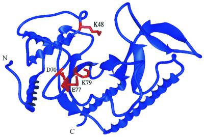
Figure 1.
Location of the mutated amino acid residues in the MutH structure. The ribbon diagram of MutH was made from the B monomer with the coordinates submitted by W. Yang (Protein Data Bank code 2AZO) using MIDAS. The residues tested from the site-directed mutagenesis, D70A, E77A and K79A, are displayed in red. An additional residue (K48) that has been shown to be involved in catalysis (12) is also displayed in red. N and C denote the N- and C-terminal ends of the protein, respectively.

