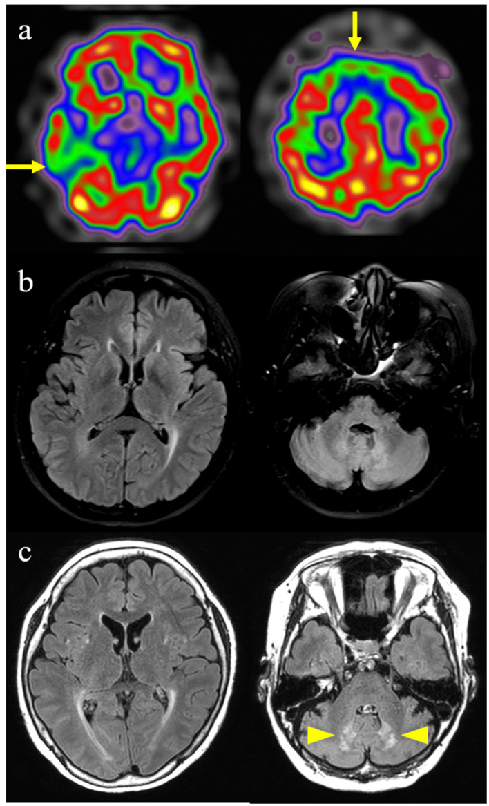
Figure 2.
Brain imaging findings of this patient with cerebrotendinous xanthomatosis. (a) Brain spectroscopy shows hypoperfusion in bilateral frontal and right temporal lobes (arrows). (b) Initial fluid-attenuated inversion recovery image shows slight periventricular white matter changes. (c) Follow-up fluid-attenuated inversion recovery image performed 4 years later shows the progression of periventricular white matter changes and newly developed symmetrical hyperintense lesions in the dentate nucleus of the cerebellum (arrowheads).

