Figure 3.
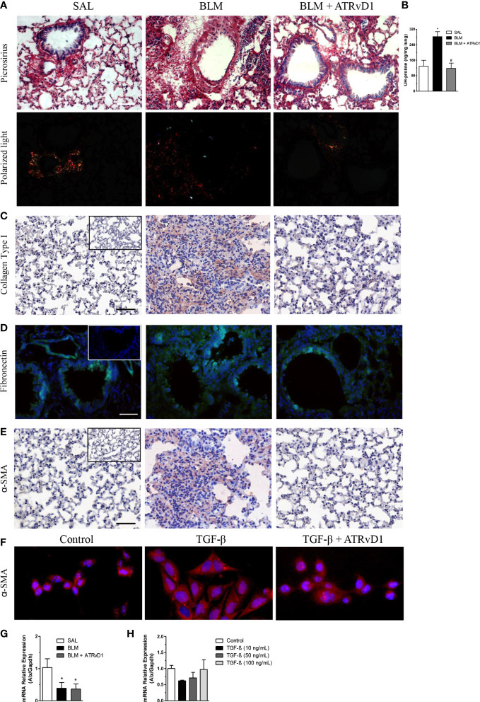
ATRvD1 impairs BLM-induced matrix protein deposition in the lung. Lungs were harvested on the 14th day after the SAL, BLM, or BLM + ATRvD1 challenges, as described in the Material and methods section. (A) Picrosirius red staining under bright field (superior images) and polarized light (lower images). (B) Hydroxyproline content in the lungs on the 14th day after mice were instilled with SAL, BLM, or BLM + ATRvD1 (top right). Results are expressed as micrograms per milligram of lung tissue. *P ≤ 0.05 compared with the SAL group; # P ≤ 0.05 compared with the BLM group. (C–E) Lung sections were immunostained for collagen type I and α-SMA by immunohistochemistry and counterstained with hematoxylin or for fibronectin and DAPI by immunofluorescence. (F) Primary lung fibroblasts were isolated, cultured, and incubated with the medium, TGF-β (10 ng/ml), or TGF-β + ATRvD1 (100 ng/ml) and then stained by immunofluorescence for α-SMA as described. Pictures are representative of each group, constituted of n = 4-5. Inserts in panels (C–E) represent the images obtained by immunostaining for the control isotype antibodies. (G, H) Total RNA was extracted from the lung tissue obtained from the SAL-, BLM-, or BLM + ATRvD1-treated mice or from the homogenate of 106 primary mouse lung fibroblasts incubated with medium or TGF-β at 10, 50, and 100 ng/ml. Samples were prepared using the TRIzol reagent and quantitative real-time RT-qPCR for ALX expression determined as described in the Material and methods section. Scale bar = 50 μm.

