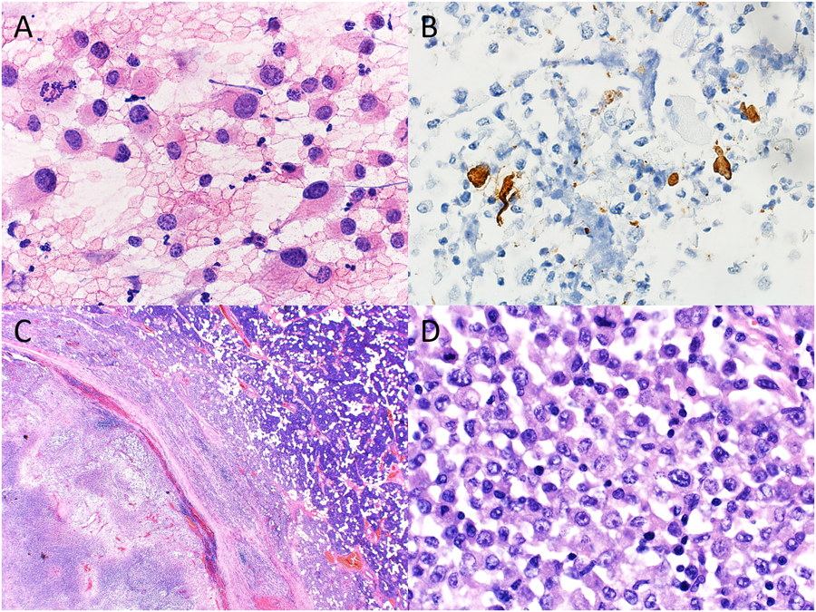
Figure 1.

Cytopathologic-histologic correlation of a case of metastatic melanoma with unknown primary site involving an intraparotid lymph node from an 18-year-old male. (A) Fine-needle aspiration (FNA) of an enlarged intraparotid lymph node demonstrated a population of large, pleomorphic cells with granular, hyperchromatic nuclei and conspicuous nucleoli (H&E, original magnification X600). (B) Immunohistochemistry performed on the cell block preparation demonstrated positivity for HMB45 in a small subset of tumor cells (immunohistochemistry, original magnification X400). (C) Surgical resection of the parotid gland showed an intraparotic lymph node with effaced architecture and extensive necrosis (H&E, original magnification X20). (D) Clusters of pleomorphic neoplastic cells with cytologic features similar to those on FNA smear were best seen in the subcapsular region (H&E, original magnification X600).
