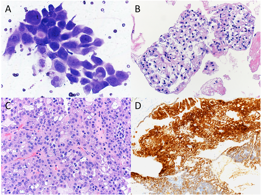
Figure 3.

Cytopathologic-histologic correlation of a case of secretory carcinoma arising in the parotid gland from a 11-year-old female. (A) Fine-needle aspiration (FNA) demonstrated frequent clusters of polygonal epithelial cells forming acinar-like structures. Tumor cells exhibited ovoid-to-round nuclei, finely granular cytoplasm with abundant small vacuoles, and occasional intracytoplasmic mucin (Diff-Quik, original magnification X600). (B) Clusters of tumor cells forming papillary structures with clear to eosinophilic cytoplasm and more frequent intracytoplasmic mucin were present in the cell block preparation (H&E, original magnification X400). (C) Surgical resection showed proliferation of bland tumor cells with papillary and acinar architectures. Intracytoplasmic eosinophilic colloid-like material was evident (H&E, original magnification X400). (D) By immunohistochemistry, the tumor cells were positive for S-100 (immunohistochemistry, original magnification X100).
