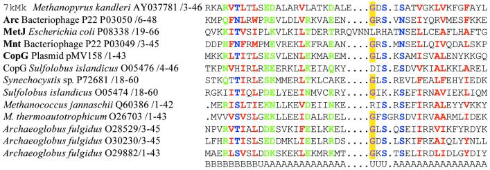
Figure 2.
Multiple alignment of 7kMk, RHH proteins with solved crystal structures (names in bold) and several putative archaeal RHH proteins. Hydrophobic (LIYFWVMA), polar (STNREQHD) and small (GASNSTCP) residues conserved in >80% of the sequences are colored red, green and blue, respectively. The conserved glycine residue of the turn connecting α-helices is colored violet and highlighted in yellow. The names of proteins, origins, accession codes and numbering of residues are indicated. Elements of secondary structure are given below the alignment. A, α-helix; B, β-strand; U, unordered.

