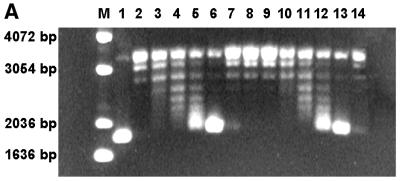
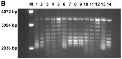
Figure 6.
DNA topology assay of 7kMk protein. Relaxed pUC19 (300 ng) was incubated with various concentrations of 7kMk in GB buffer at 70°C for 30 min. 7kMk was added at a Rw of 0 (lane 10), 0.4 (lane 3), 0.8 (lanes 4 and 11), 1.6 (lanes 5 and 12), 3.3 (lanes 6 and 13) 5 (lane 7), 6.7 (lanes 8 and 14) and 12 (lane 9). The resulting complexes were digested with topoisomerase V (100 ng) at the same temperature for 15 min (lanes 3–10) or 1 h (lanes 11–14). Lanes M, 1 and 2 were a 1 kb DNA ladder (Gibco BRL), negatively supercoiled pUC19 DNA isolated from E.coli and relaxed pUC19 DNA, respectively. The reaction products were analyzed by 1.5% agarose gel electrophoresis in TBE buffer (A) or TBE buffer containing 1 µg/ml chloroquine (B) at 1.5 V/cm for 16 h, stained with ethidium bromide and digitized.


