Fig. 3.
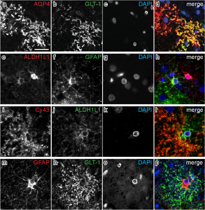
Double-labelled immunofluorescence shows co-expression of different astrocyte markers. Each horizontal set comprises images taken from the same field of view, showing immunostaining for different markers in the superior frontal cortex. The colour of each label in the merged image is represented by the colour of the text in the relevant monochrome image. Sections were counterstained with DAPI to identify nuclei. a–d. Aquaporin-4 (AQP4) and glutamate transporter 1 (GLT-1) both labelled astrocytic processes and end feet, and are a largely overlapping population of astrocytes. e–h. Aldehyde dehydrogenase-1 L1 (ALDH1L1) and glial fibrillary acidic protein (GFAP) label the cell body and processes of astrocytes. i–l. An astrocyte immunolabelled with connexin-43 (Cx43) and ALDH1L1. m–p. Different cellular compartments are labelled with GFAP and GLT-1, and are largely a non-overlapping population of astrocytes. Scale bar in a = 20 μm (applies to all images)

