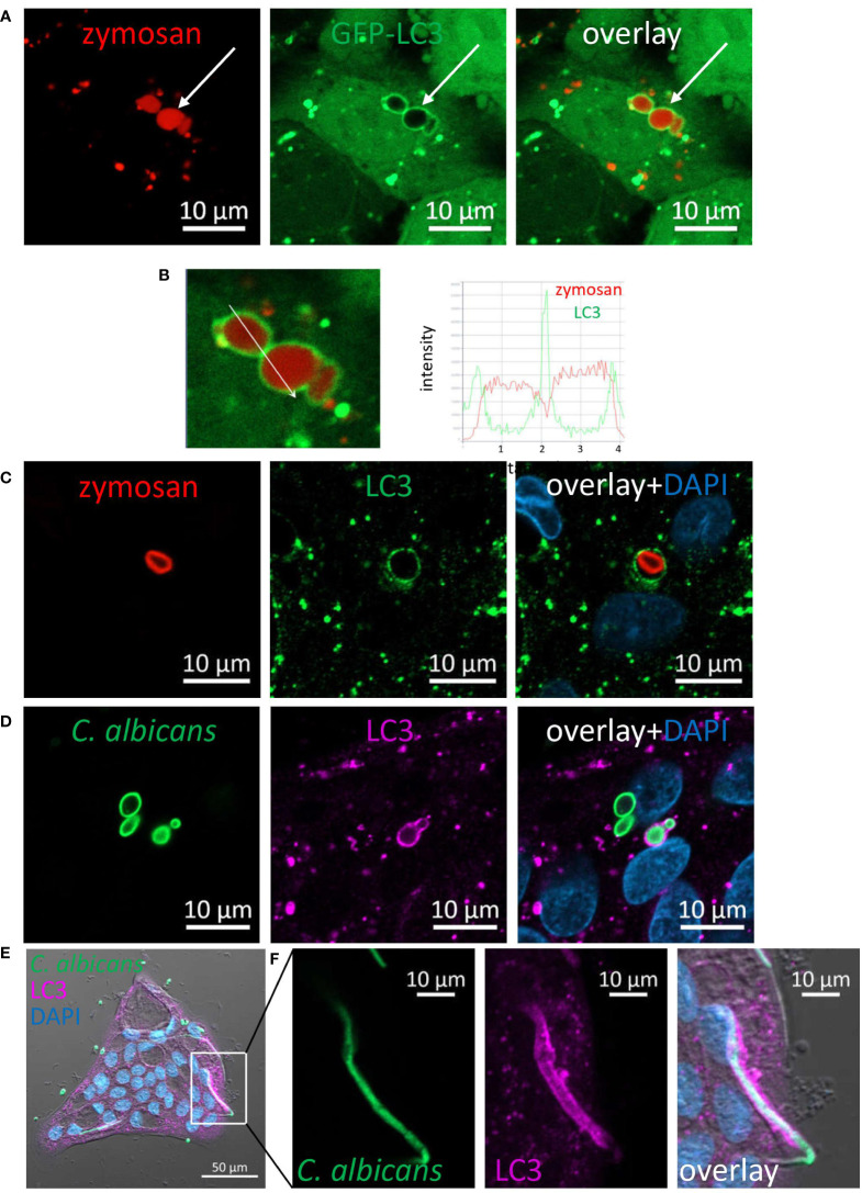
Figure 4.
LC3 is recruited to phagosomes in IECs. (A) SW480 LC3-GFP cells were fed with pHrodo-red zymosan overnight. Live imaging shows LAPosomes (arrow) as LC3 (green) around intact zymosan (red) particles, as well as fragmented zymosan and autophagosomes. (B) Fluorescence intensity profile along the arrow of an inset from (A) is shown on the graph. (C) Colonic organoids were fed with pHrodo-red zymosan (red) overnight and stained with LC3 antibody (green) and DAPI (blue). (D–F) Colonic organoids were fed with Rhodamine-green-X HK-C. albicans (green) overnight and stained with LC3 antibody (magenta) and DAPI (blue). Shown is LAP of yeast (D) and hyphal form (E, F) of HK-C. albicans. F is an inset of (E) Original magnification ×40 (A, B), ×63 (C, D) x20 (E, F) scale bar 10 µm (A–D, F) and 50 µm (E).

