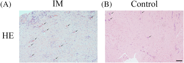
FIGURE 2.

Haematoxylin and eosin (HE) staining of the induced membrane (IM) and the control periosteal membrane. A, The intensely fibrous, cell‐rich, vascularised tissue of the IM. B, Very few vessel‐like structures seen in the normal periosteum. Black arrows indicate new blood vessels. Scale bar: 500 μm
