
Figure 3.
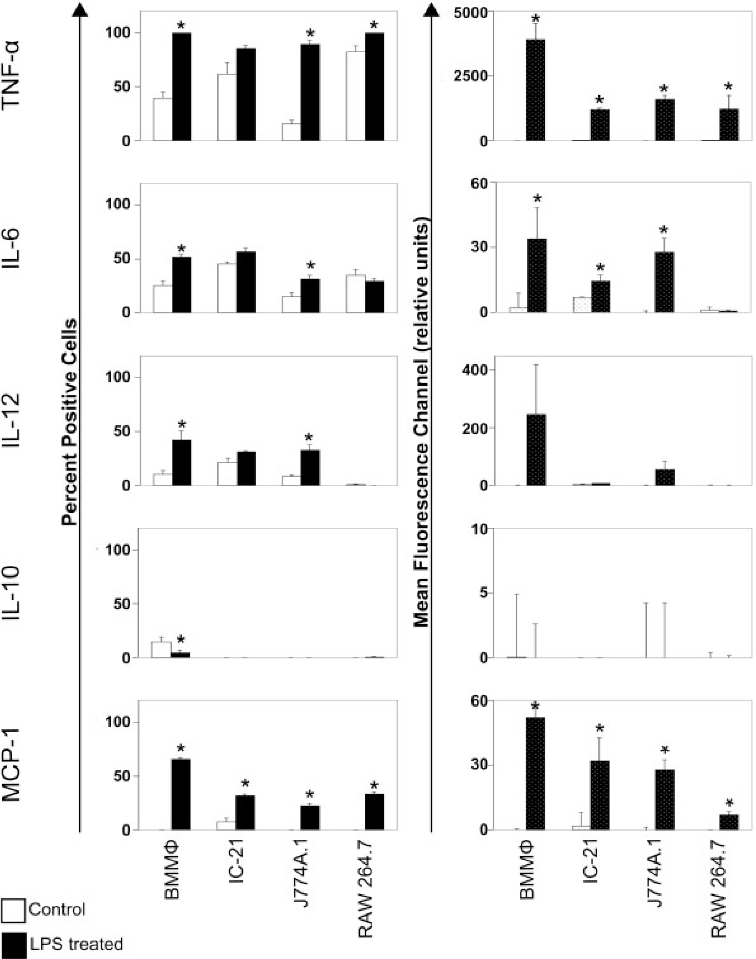
Flow cytometric analysis of inflammatory cytokines. Cells were incubated with LPS and/or a complex to stop Golgi complex export of cytokines for 8 h prior to intracellular immunostaining and flow cytometry analysis. White bars indicate basal cytokine expression on TCPS and black bars indicate cytokine expression after treatment with LPS. Error bars represent standard error. The induction of cytokine expression varies greatly from cell type to cell type. (Data are representative of at least three experiments, statistical significance of p ≤ 0.05 is indicated by an asterisk).