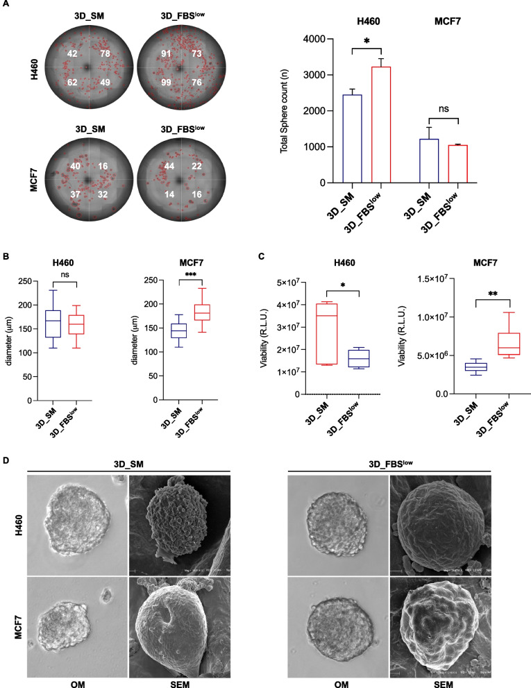
Fig. 5.
Analysis of morphology and growth rate of H460- and MCF7-derived tumor spheroids. A Representative images and relative histograms of H460 3D_SM, H460 3D_ FBSlow, MCF7 3D_SM, and MCF7 3D_FBSlow tumor spheroids morphology, count and B diameter. C Cell viability of H460 3D_SM, H460 3D_FBSlow, MCF7 3D_SM, and MCF7 3D_FBSlow assessed by Cell titer-Glo 3D assay and expressed as relative light unit (RLU). D Representative images of H460 3D_SM, H460 3D_FBSlow, MCF7 3D_SM, and MCF7 3D_FBSlow tumor spheroids obtained by SEM. All the experiments were carried out in triplicate and results are presented as mean ± SD. p-value: * < 0.05, ** < 0.01. *** < 0.001. ns: not significant

