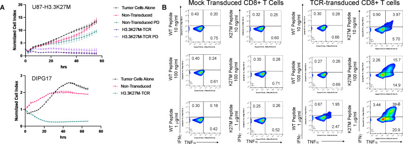
Figure 1.
Functions of H3.3K27M-TCR-transduced T cells. (A) Cytotoxicity assays using a live cell imaging system. U87-H3.3K27M (upper panel) or HSJD-DIPG-0172 (lower panel) were seeded on laminin-coated plates and were rested for 24 hours to attain optimum cell index. Effector CD8+T cells were added at an E:T ratio of 5:1. U87-H3.3K27M cells (upper panel), were in the assay with control, non-transduced but similarly activated CD8+T cells were included. ‘PD’ refers to ‘process development’, because these cells were derived from GMP-grade manufacturing test cultures of the cells using Miltenyi’s Prodigy system. Cytotoxicity against glioma cells was monitored for 56 (upper panel) or 66 (lower panel) hours using the xCELLigence platform (Agilent). The experiment was performed at least twice with similar results and graphs represent means of triplicate cultures±SD. (B) Intracellular cytokine expression assays. T2 cells were plated at a density of 50,000 cells/well in 100 µL with the indicated concentrations of WT or H3.3K27M peptide. Aliquots of 50,000 control or TCR-transduced CD8+ cells were added to each well in 100 µL. Cells were harvested after 5 hours and stained for intracellular markers. TCR, T cell receptor; WT, wild-type.

