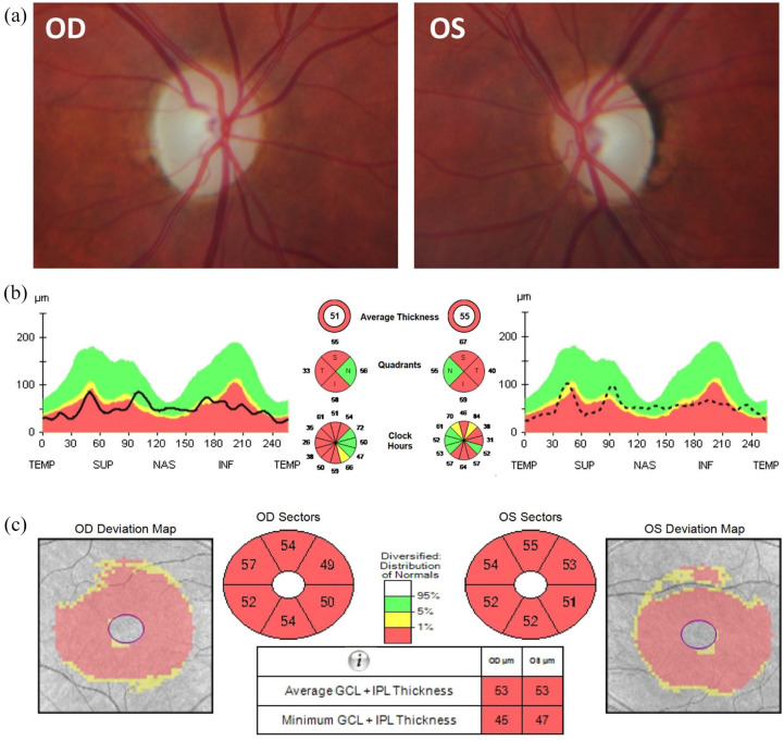
Figure 1.
Ophthalmological imaging findings in a patient with WFS1 spectrum disease. A 32-year-old woman developed progressive bilateral visual loss from the age of 27 years. She did not have a history of deafness, diabetes mellitus, or diabetes insipidus. There was no family history of early-onset progressive visual loss. Magnetic resonance imaging revealed small optic nerves without abnormal contrast enhancement. Whole mitochondrial genome sequencing did not detect any pathogenic variants, including the three most common mtDNA mutations associated with LHON. However, she was found to carry compound heterozygous WFS1mutations (c.605A >G p.(Glu202Gly); c.874C >T p.(Pro292Ser)). On examination, the best-corrected visual acuity was 6/24 in both eyes. Color vision was 9/15 Ishihara plates in the right eye (OD) and 7/15 in the left eye (OS). There was no relative afferent pupillary defect. (a) Both optic nerves were pale. (b) OCT imaging of the optic nerves revealed significant retinal nerve fibre layer loss with some sparing of the nasal fibers bilaterally. (c) OCT of the macula showed symmetric generalized atrophy of the GCL and IPL in both eyes.
GCL, ganglion cell layer; IPL, inner plexiform layer; INF, inferior; LHON, Leber hereditary optic neuropathy; mtDNA, mitochondrial DNA; NAS, nasal; OCT, optical coherence tomography; OD, oculus dextrus; OS, oculus sinister; SUP, superior; TEMP, temporal.

