. 2023 Feb 22;12:e78512. doi: 10.7554/eLife.78512

Figure 1. Establishing intersectional tools for genetic access to enteroendocrine cells in vivo.

(A) Bright-field microscopy and native GFP fluorescence microscopy of intestinal tissue from Vil1-p2a-FlpO; fsf-Gfp mice (left) and fsf-Gfp mice (right). Scale bars: 5 mm. (B) Cartoon depicting intersectional genetic strategy to access enteroendocrine cells. (C) Native reporter fluorescence in cryosections (20 μm, except 50 μm for cortex and dorsal root ganglion) or wholemounts (tongue) of fixed tissues indicated from Neurod1-Cre; lsl-tdTomato mice (left), Vil1-p2a-FlpO; fsf-Gfp mice (middle), and Neurod1INTER; inter-tdTomato mice (right). Scale bars: 100 μm for all except 500 μm for tongue. Intestine sections from duodenum (middle) or jejunum (left, right). See Figure 1—figure supplement 1.

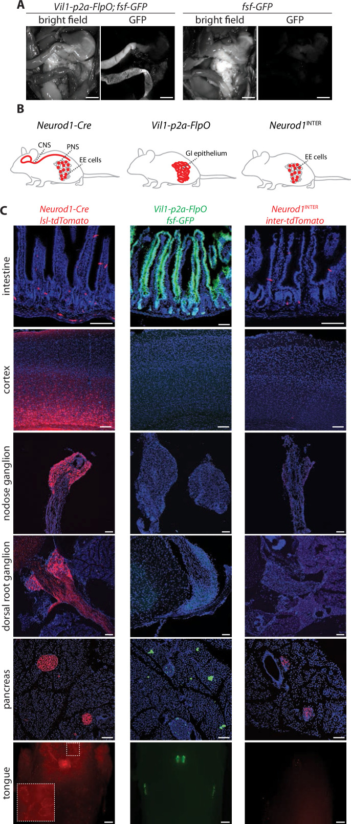
Figure 1.

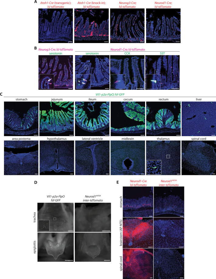
Figure 1—figure supplement 1. Characterization of mouse lines for intersectional genetics.

Figure 1—figure supplement 1.

(A) Native tdTomato fluorescence in fixed intestinal cryosections (20 μm) of mouse lines indicated. (B) Two-color analysis examining expression of tdTomato (native fluorescence, magenta) and hormones (immunochemistry, green) in fixed intestinal cryosections (20 μm) of mouse lines indicated, SST: somatostatin. (C) Native GFP fluorescence in fixed cryosections of tissues (20 μm), spinal cord (50 μm) and brain regions indicated (50 μm) in Vil1-p2a-FlpO; fsf-Gfp mice. (D) Native reporter fluorescence in fixed wholemount tissue preparations from mouse lines indicated. Scale bars: 500 μm. (E) Native tdTomato fluorescence in fixed tissue cryosections (20 μm for stomach, 50 μm for cortex, spinal cord) from mouse lines indicated. Scale bars: 100 μm.