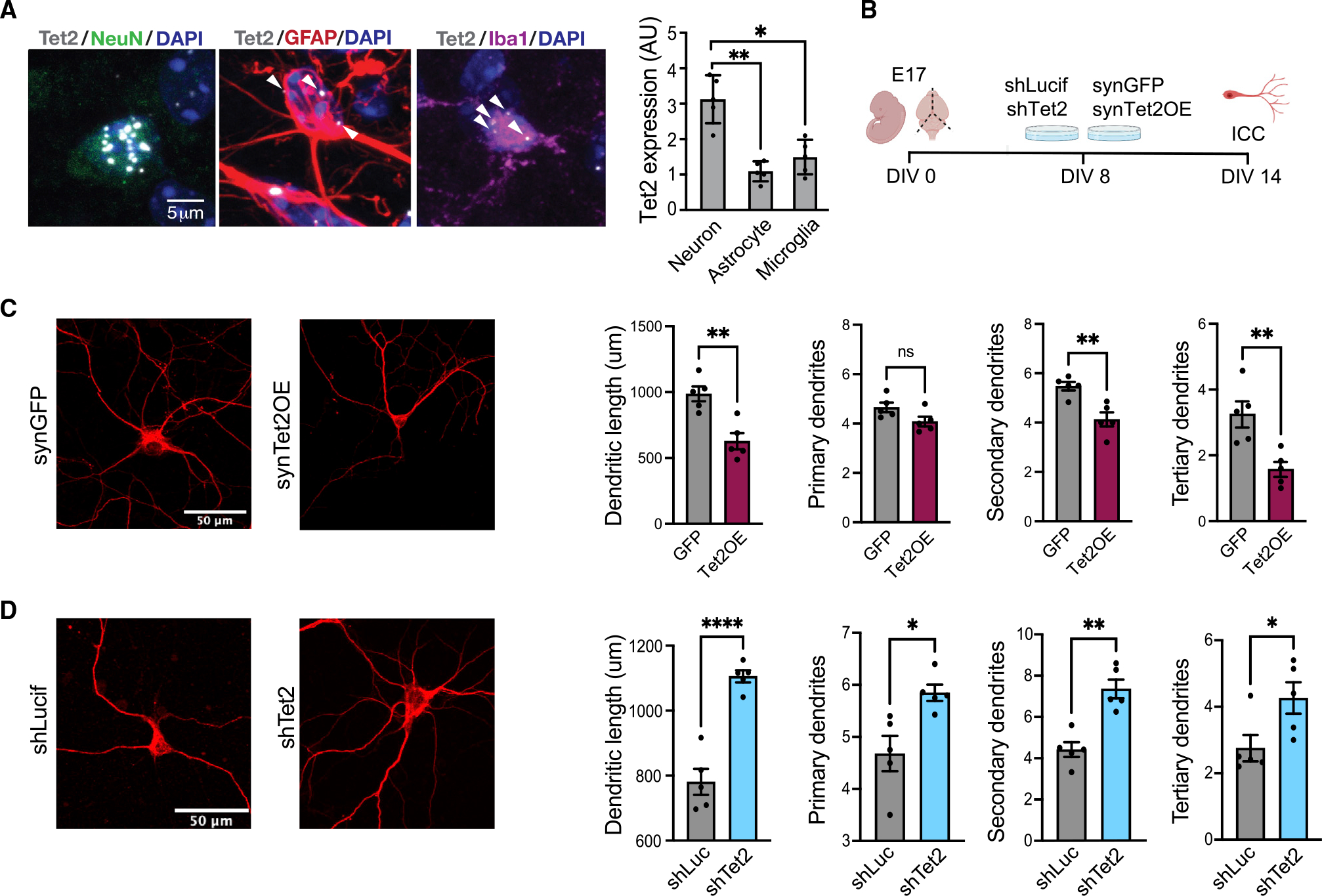
Figure 1. Tet2 is enriched in neurons and regulates neuronal complexity.

(A) Representative image and quantification of Tet2 RNA scope expression in hippocampal neurons, astrocytes and microglia of young (3 months old) adult mice (n = 5 mice with 20–40 cells per animal; one-way ANOVA; *p < 0.05, **p < 0.01).
(B) Schematic of experimental design. Primary mouse neurons were cultured for 8 days and subsequently infected with lentivirus encoding Tet2 (Tet2 OE) or GFP control sequences under the neuron-specific Synapsin1 promoter or encoding shRNA sequences targeting Tet2 (shTet2) or luciferase control (shCtrl).
(C) Representative images and quantification of dendritic length and number of primary, secondary, and tertiary dendrites for MAP2-postive neurons following viral-mediated Tet2 overexpression (n = 5 per group; t test; *p < 0.05, **p < 0.01).
(D) Representative images and quantification of dendritic length and number of primary, secondary, and tertiary dendrites for MAP2-postive neurons following viral-mediated Tet2 knockdown (n = 5 per group; t test; *p < 0.05, **p < 0.01, ****p < 0.0001).
Data are represented as mean ± SEM.
See also Figure S1.
