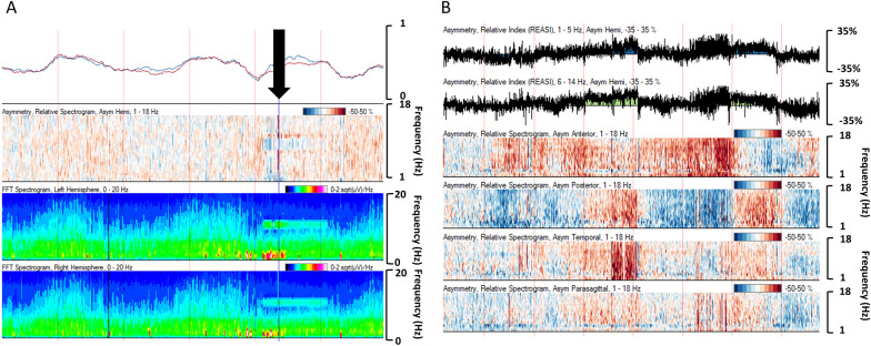
Fig. 9.
a Artifact in the 13- to 14-Hz frequency range from a high-frequency chest wall oscillation vest (black arrow) in a 13-year-old. b Quantitative electroencephalography (EEG) in a 3-month-old with congenital heart disease status post surgical repair. Asymmetry indices and spectrograms demonstrate shifting impact of scalp edema on the EEG signal, with changing asymmetric power at regular intervals, reflecting timed repositioning of the patient by the bedside nurses

